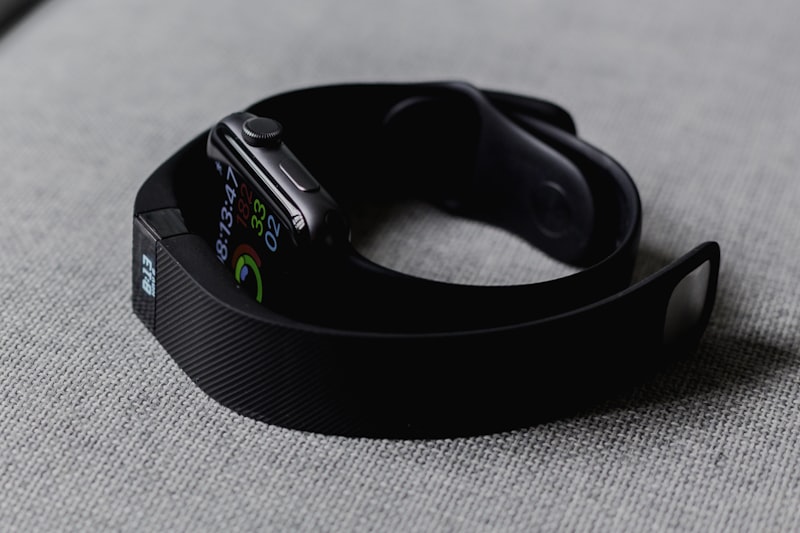
Every wrist-worn health tracker rests on a deceptively simple idea: shine light into the skin, and read what comes back. The blood pulsing through the vessels beneath your wrist subtly changes how much light returns, and from that flicker a modern wearable reconstructs your heart rate, the spacing between beats, your blood oxygen and more. This is photoplethysmography, and understanding it clarifies both what these devices can tell you and where their limits lie.
The term photoplethysmography — usually shortened to PPG — combines the Greek roots for light, volume and writing. Literally, it means writing a record of volume changes using light. That volume is blood, and the changes are the rhythmic swelling and relaxation of the small vessels in your skin with every heartbeat. A PPG sensor does not measure the heartbeat directly. It measures the optical shadow that circulating blood casts, then infers the mechanics of the cardiovascular system from that shadow.
What makes this remarkable is that the underlying hardware is modest: a few light-emitting diodes, a light-sensitive detector, and a great deal of signal processing. The sophistication lives not in exotic components but in the mathematics that separates a faint, meaningful pulse from a torrent of noise. To appreciate a wrist sensor honestly, it helps to follow the light on its short journey into the skin and back.
1. Light, Blood and the Basic Principle
Human tissue is partly translucent. Shine a bright light against your fingertip and you will see a reddish glow through the other side — light is passing through skin, muscle and blood, and being selectively absorbed along the way. Blood, and specifically the hemoglobin that carries oxygen, absorbs certain wavelengths of light more strongly than the surrounding tissue does. This selective absorption is the physical hook on which all PPG depends.
When your heart contracts, a pulse of blood surges into the arteries and the small vessels of the skin. For a brief moment there is slightly more blood in the illuminated patch of tissue, so slightly more light is absorbed and slightly less returns to the detector. When the heart relaxes, blood volume ebbs and more light returns. The detector therefore sees a signal that rises and falls in step with the pulse — a waveform whose peaks and troughs trace the cardiac cycle.
The AC and DC components
Engineers describe this signal as having two parts. The large, slowly changing background — absorption by skin, bone, tissue and the non-pulsing blood — is called the DC component. Riding on top of it is a small, rhythmic fluctuation caused by the pulsing arterial blood, called the AC component. The AC portion is tiny, often well under a few percent of the total signal, yet it carries nearly all the useful information. Extracting that small oscillation cleanly from the large steady background is the central engineering task of every wrist sensor.
A PPG sensor never measures your heartbeat directly. It measures how the volume of blood beneath the skin changes the amount of light returning to a detector — and reconstructs the beating heart from that faint optical rhythm.
2. Why Green Light for Heart Rate
Most wrist wearables use green light for everyday heart-rate tracking, and the choice is deliberate. Hemoglobin absorbs green light strongly, which means the pulsing blood produces a comparatively large change in the returning green signal. A larger change is easier to detect against noise, so green light yields a robust, high-contrast pulse waveform — exactly what you want when the wearer is walking, typing or exercising.
Green light also penetrates only shallowly into tissue. That sounds like a disadvantage, but at the wrist it is a strength: it confines the measurement to the superficial vessels and is less easily disturbed by deeper motion. In bright daylight and during movement — the very conditions in which reliable heart rate matters most — green light gives wrist devices their best signal-to-noise ratio.
The trade-off
The cost is that green light is not well suited to measuring blood oxygen. Estimating oxygen saturation requires comparing how blood absorbs two different wavelengths, and for that comparison to work the wavelengths must sit where oxygenated and deoxygenated hemoglobin behave distinctly. Green does not satisfy that condition well. So devices that measure oxygen carry additional LEDs of other colours, and switch between them depending on what they are trying to learn.
3. Red and Infrared for Blood Oxygen
Pulse oximetry — the estimation of blood oxygen saturation, often written SpO2 — rests on a specific optical fact. Oxygenated hemoglobin and deoxygenated hemoglobin absorb red and infrared light in opposite ways. Oxygen-rich blood lets more red light through and absorbs more infrared; oxygen-poor blood does the reverse. By shining both red and infrared light into the skin and comparing the pulsing signals each produces, a device can estimate the ratio of oxygenated to total hemoglobin.
The method works on the pulsatile, arterial part of the signal specifically. Because the AC component comes from arterial blood surging with each beat, comparing the red and infrared AC signals isolates the property of arterial blood — which is what oxygen saturation is meant to describe. The device computes a ratio of ratios and maps it, through a calibration curve, to an estimated saturation percentage.
Green light is chosen for its strong, motion-resistant pulse signal; red and infrared are chosen because oxygenated and deoxygenated blood absorb them in opposite ways. Different questions call for different colours of light.
Wrist-based oximetry is harder than the fingertip clip used in hospitals. At the fingertip, light passes through thin, well-perfused tissue with vessels close to the surface. At the wrist, the sensor works in reflection — light goes in and bounces back from the same side — through tissue that is thicker and less richly supplied with blood. This is one reason wrist SpO2 readings are best treated as trend indicators for wellness rather than clinical-grade measurements.
4. From Waveform to Heart Rate and HRV
Once a clean pulse waveform exists, deriving heart rate is conceptually straightforward: count the peaks over time. Each peak marks one cardiac cycle, and the number of peaks per minute is the heart rate. The real subtlety lies in measuring the interval between consecutive beats with enough precision to study its variation.
The spacing between successive heartbeats is never perfectly constant. It lengthens and shortens by small amounts, breath by breath and moment by moment, under the influence of the autonomic nervous system. This beat-to-beat variation is heart-rate variability, or HRV, and it has become one of the most watched signals in consumer wellness because it offers an indirect window onto the balance between the body's stress and recovery systems.
The precision problem
Measuring HRV from PPG demands timing accuracy on the order of milliseconds, because the variations being studied are themselves small. A clinical electrocardiogram detects the sharp electrical spike of each heartbeat and can time beats very precisely. PPG instead detects a smooth, rounded optical pulse whose peak is inherently less crisp, and which arrives slightly after the electrical event as the pressure wave travels to the wrist. Careful devices therefore measure HRV during still periods — typically overnight — when motion is minimal and the pulse shape is most stable, and they treat the result as a well-correlated proxy rather than an identical substitute for electrocardiogram-derived variability.
5. The Signal-Processing Challenge
The clean waveform described so far is an idealisation. In the real world the raw optical signal is buried under interference, and recovering a trustworthy pulse is the hardest part of the whole enterprise. Three sources of trouble dominate.
Motion, ambient light and perfusion
Motion is the largest. When the wrist moves, the sensor shifts against the skin, tissue compresses, and blood sloshes in ways unrelated to the heartbeat. These movements can produce fluctuations far larger than the true pulse, and worse, rhythmic motion such as running can masquerade as a plausible heart rhythm. Ambient light is the second: sunlight or lamplight leaking into the detector adds a spurious signal that must be rejected. The third is low perfusion — when the skin is cold, or the vessels are constricted, there is simply less pulsing blood to modulate the light, and the signal grows faint.
To fight these, devices combine optical and computational defences. Sensor housings are designed to block stray light and maintain steady contact. Detectors sample the signal with the LEDs briefly off to measure and subtract ambient light. And crucially, a separate motion sensor — an accelerometer — records how the wrist is moving, so algorithms can recognise and remove the motion-induced components from the optical signal. This last technique, using the accelerometer as a reference for what to cancel, is one of the most important advances in wrist-based sensing.
6. Accuracy, Skin Tone and Honest Limits
No account of PPG is complete without a frank discussion of accuracy. These sensors are wellness instruments, not medical devices, and the distinction is not marketing caution — it reflects genuine physical constraints.
One well-documented consideration is skin tone. Melanin, the pigment that gives skin its colour, absorbs light — and it absorbs green and, to a lesser degree, red light. In more pigmented skin, more of the emitted light is absorbed by melanin before it ever reaches the blood vessels, weakening the pulse signal that returns to the detector. Studies have found that this can reduce signal quality and, in some conditions, affect the reliability of readings across skin tones. Responsible manufacturers address this with brighter and more varied light sources, multiple detectors and adaptive algorithms, but it remains a real factor that the field continues to work on, and honesty about it matters.
Wellness versus clinical measurement
Other limits are inherent. Wrist perfusion varies with temperature and posture. Tattoos over the sensor can absorb light unpredictably. A loose fit lets in stray light and motion. Even under good conditions, a reflective wrist reading cannot match the accuracy of a transmissive fingertip clip or a chest electrocardiogram. This is why a well-designed device frames its numbers as trends and relative changes rather than diagnostic absolutes — a resting heart rate that is rising over weeks is meaningful even if any single reading carries uncertainty.
The most useful thing a wrist sensor offers is not a single perfect number but a long, consistent record of your own patterns. A measurement that is slightly imprecise but taken thousands of times, always on the same body, reveals change that no one-off reading can.
7. Turning Signals into Insight
An individual PPG reading, however clean, is only a data point. The value emerges when many signals are fused and observed over time. This is where sensing becomes insight.
Consider sleep. No single sensor detects sleep directly. Instead, a device blends the slowing and steadying of heart rate, the rise in heart-rate variability that accompanies rest, the stillness reported by the accelerometer, subtle shifts in skin temperature and respiratory rate inferred from the way breathing modulates the pulse, all set against the time of night. Taken together, these signals let algorithms estimate not just whether you are asleep but the texture of that sleep. The same fusion underpins measures of recovery and daily readiness: how far tonight's resting heart rate and variability sit from your own normal tells a richer story than any figure in isolation.
The essential ingredient is a personal baseline. Because healthy values differ enormously between individuals, what matters is not how your numbers compare to a population but how today compares to your own established pattern. A device like Aura Clarus builds this longitudinal baseline quietly over weeks of wear, learning the range that is normal for you so that a meaningful departure can be recognised as such. That patient accumulation — the same body measured again and again — is what converts a stream of imperfect optical readings into a signal worth attending to.
8. Where the Wrist Goes Next
Photoplethysmography has already travelled a long way from its origins as a laboratory curiosity to a sensor worn by hundreds of millions. Its future is likely to be one of refinement and combination rather than a single dramatic leap. Additional wavelengths and multiple detectors promise better performance across skin tones and conditions. Smarter algorithms, increasingly running on the device itself, will separate signal from noise more effectively and extract information — such as respiratory patterns and estimates of vascular behaviour — that today is only beginning to be read from the same optical stream.
The broader trajectory is toward context. A raw number means little; the same number interpreted against your history, your activity and your other signals can mean a great deal. As wearables grow better at that interpretation, and as they pair sensing with thoughtful guidance, their role shifts from displaying data to supporting understanding. The physics at the heart of it remains what it always was — light into skin, light back out, and blood volume written in the difference. What changes is how carefully, and how honestly, we learn to read it.
Understood for what it is, the wrist sensor is a quietly impressive piece of engineering: a way to glimpse the beating cardiovascular system through the skin, without a needle or a wire, thousands of times a day. It will not replace a clinic. But as a patient, personal observer of your own patterns, it offers something a clinic cannot — continuity — and that is worth taking seriously, limits and all.
This article is published by Adarna Inc. for educational and informational purposes only. It does not constitute medical advice. Consult a qualified healthcare professional before making any health-related decisions.